Home
/ Human Back Bones Diagram - Spine Anatomy Overview Video : It also covers some common conditions and injuries that can affect the back.
Human Back Bones Diagram - Spine Anatomy Overview Video : It also covers some common conditions and injuries that can affect the back.
Human Back Bones Diagram - Spine Anatomy Overview Video : It also covers some common conditions and injuries that can affect the back.. Just need a glimpse, leave your valuable advice let us know , and subscribe us! It also covers some common conditions and injuries that can affect the back. The notochord present in the embryonic stage is replaced by the vertebral column. The lumbar spine connects to the thoracic spine above and the hips below. At birth, the skeleton of a newborn has more than 300 bones;
Long, short, irregular, and flat. The occiput (co), also known as the occipital bone, is a flat bone that forms the back of the head. Check out our bones diagram selection for the very. And coccygeal the tail bone. It also covers some common conditions and injuries that can affect the back.
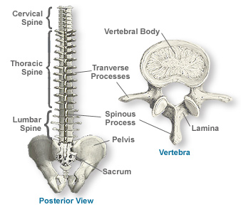
Anatomy Of The Spine Southern California Orthopedic Institute from www.scoi.com The vertebral column houses the spinal canal, a cavity that. Lateral labeled diagram of the human vertebral spinal column showing vertebrae numbering order and the 5 different regions of the spine. Skeletal system diagrams are illustrations of the human skeleton, used mostly for educational purposes or in presentations. It contains the osteology, arthrology and myology of the spine and back. Human back bones diagram poster 28 inch x 24 inch 16 inch x 13 inch. These bones are connected at the back with specialized joints. Spinal vertebrae bone spine vertebra toracica spinal cord spine structure back diagram spine sections spinal cord vertebrae spinal structure health diagram. Skeleton back bones diagram / human skeleton anatomy vintage 1940s high res digital image / in this assignment, students color the various parts of the skeletal system and then answer some follow up teach your students the names of the bones in the human body with the help of this illustrated human skeleton diagram.
There are four main categories of bones:
Its appearance is different from the other spinal vertebrae. Human back bones diagram poster 28 inch x 24 inch 16 inch x 13 inch. This vertebra supports the skull. Bone name diagram catalogue of schemas. The atlas is the topmost vertebra, and along with c2, forms the joint connecting the skull and spine. Flat bones follow the process of intramembranous ossification where the young bones grow from a primary ossification center in fibrous membranes and leave a small region of. A human pelvis is much wider from the side with curved bones. This human anatomy module is composed of diagrams, illustrations and 3d views of the back, cervical, thoracic and lumbar spinal areas as well as the various vertebrae. Diagram of a human female skeleton, back view. Vertebrae separated by intervertebral discs. We are pleased to provide you with the picture named anatomy of back muscles diagram.we hope this picture anatomy of back muscles diagram can help you study and research. Skeletal diagrams are tools used by students to learn and study all 206 bones (this number can vary from person to person) of the human body. 12 photos of the human back bone chart.
This human anatomy module is composed of diagrams, illustrations and 3d views of the back, cervical, thoracic and lumbar spinal areas as well as the various vertebrae. The notochord present in the embryonic stage is replaced by the vertebral column. The human body is an incredible machine. The vertebral column, also known as the backbone or spine, is part of the axial skeleton.the vertebral column is the defining characteristic of a vertebrate in which the notochord (a flexible rod of uniform composition) found in all chordates has been replaced by a segmented series of bone: Diagram of a human female skeleton, back view.
Anatomy Of The Back Spine And Back Muscles Kenhub from thumbor.kenhub.com Our latest youtube film is ready to run. But, they are common in the back and can cause pain. It contains the osteology, arthrology and myology of the spine and back. The first seven bones (vertebrae) of your spine form your neck. 12 photos of the human back bone chart. Human back bones diagram poster 28 inch x 24 inch 16 inch x 13 inch. The movable vertebrae are divided… read more A tough, springy disc of cartilage sits between the vertebrae of your spine.
This vertebra supports the skull.
The spine anatomy is a complex structure. The spine diagram the spine diagram shown below, consists of many bones or vertebrae,soft discs,the spinal cord, and spinal nerves. In the back and elsewhere in the body, tendons attach muscles to bones. Human backbone diagram, bone, human backbone diagram. The vertebral column is a part of the axial skeleton, which comprises the skull, ribs and sternum other than the vertebral column. Long, short, irregular, and flat. As a person ages, these bones grow together and fuse into larger bones, leaving adults with only 206 bones. When autocomplete results are available use up and down arrows to review and enter to select. The red lines point individual bones and the names are writen in singular, the blue lines conect to group of bones and are in plural form. Muscles of the abdomen lower back and pelvis. The atlas is the topmost vertebra, and along with c2, forms the joint connecting the skull and spine. The first seven bones (vertebrae) of your spine form your neck. Flat bones follow the process of intramembranous ossification where the young bones grow from a primary ossification center in fibrous membranes and leave a small region of.
This article looks at the anatomy of the back, including bones, muscles, and nerves. And coccygeal the tail bone. At birth, the skeleton of a newborn has more than 300 bones; When autocomplete results are available use up and down arrows to review and enter to select. Can you feel the bumps of your vertebrae along your back?
Lumbar Wikipedia from upload.wikimedia.org Human backbone diagram, bone, human backbone diagram. It is particularly interesting for physiotherapists. The spine or backbone consists of 26 small bones or vertebrae. Human back bones diagram poster 28 inch x 24 inch 16 inch x 13 inch. The neck (cervical) and low back (lumbar) regions have a slight concave curve, and the thoracic and sacral regions have a gentle convex curve (fig. Skeletal system diagrams are illustrations of the human skeleton, used mostly for educational purposes or in presentations. The red lines point individual bones and the names are writen in singular, the blue lines conect to group of bones and are in plural form. Muscle or tendon injuries can occur anywhere in the body.
Human body anatomy female female anatomy muscle shoulder blade pain anatomy back muscles bones man female anatomy body muscles in a body female anatomy muscole shoulder concept muscular sysyem.
These bones are connected at the back with specialized joints. The human backbone is a column of 33 total vertebrae, of which 24 are movable and free (the remainder are fused). The bones of the pelvis and lower back work together to support the body's weight, anchor the abdominal and hip muscles, and protect the delicate vital organs of the vertebral and abdominopelvic cavities. Diagram of a human female skeleton, back view. Human backbone diagram, bone, human backbone diagram. And coccygeal the tail bone. But, they are common in the back and can cause pain. Skeleton back bones diagram / human skeleton anatomy vintage 1940s high res digital image / in this assignment, students color the various parts of the skeletal system and then answer some follow up teach your students the names of the bones in the human body with the help of this illustrated human skeleton diagram. They help support particular bones and make them move. Skeletal diagrams are tools used by students to learn and study all 206 bones (this number can vary from person to person) of the human body. There are four main categories of bones: The vertebral column of the lower back includes the five lumbar vertebrae, the sacrum, and the coccyx. Skeletal system diagrams are illustrations of the human skeleton, used mostly for educational purposes or in presentations.
At the back of each bone in the spine (vertebra) are bony points called processes, which muscles attach to human back bones. Vertebrae separated by intervertebral discs.